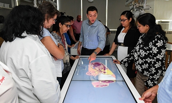
Students in the College of Pharmacy and Health Sciences will gain a deeper understanding of gross human anatomy, thanks to the installation of new Anatomage virtual dissection tables at St. John’s University’s Queens campus and the Dr. Andrew J. Bartilucci Center.
“We wanted to integrate new technologies for our current students while using the tables as a recruitment tool for prospective students,” said Sawanee Khongsawatwaja, Associate Dean, Administration and Fiscal Affairs, College of Pharmacy and Health Sciences. “The investment in these tables will help to ensure student success by enhancing the teaching and learning environment at the University.”
The Anatomage Table is the world’s first, and most technologically advanced, virtual dissection table for anatomy education. Used by leading medical schools and institutions around the world, the table can effectively replace expensive and cumbersome cadaver laboratories.
"By including real-life, 3-D projections on the Anatomage Table, we take another step forward in providing schools with the most advanced medical school-level content without the cost of dissection labs," said Jack Choi, Chief Executive Officer, Anatomage.
The 84-inch-long table features a high-resolution touch screen that allows users the ability to visualize human anatomy and patient data—exactly as they would on a real cadaver. Each anatomical structure is accurately reconstructed in 3-D and is sourced from actual human cadavers. Because of the high accuracy of the renderings, which are life-sized and true to color, the table allows for exploration and learning of human anatomy beyond what any cadaver could offer.
The virtual cadaver can be viewed and manipulated from an infinite combination of angles and zoom levels with the stroke of a finger or stylus. A virtual scalpel feature allows users to slice through tissue and bone to explore further.
These are the University’s second and third Anatomage Tables. In 2016, the first table was installed at the nearby Dr. Andrew J. Bartilucci Center. That unit was recently replaced with the same model just installed in St. Albert Hall.
In July, faculty attended day-long, on-campus training exercises with a representative from the company. There, each had the opportunity to discover the capabilities of the tables through hands-on learning exercises.
Marc E. Gillespie, Ph.D., Professor, Pharmaceutical Sciences, is among the faculty who attended the session and was impressed with the promise the new technology offers. “The operating table format presents students with a spectacularly detailed representation of a human cadaver,” he said. “This 3-D representation can be moved in real time, manipulated by students, and used for active learning and exploration.”
Each table has the capability to present four gross anatomy cases, over 20 high-resolution regional anatomy cases, and more than 1,000 pathological examples. As a result, instructors are better able to draw students’ interest and attention, which leads to more effective educational outcomes.
Initially, students enrolled in the Doctor of Pharmacy Degree program, the Physician Assistant program, and the Radiologic Sciences program will benefit from the presence of the tables on campus.
“Physician Assistant students have consistently voiced a desire to participate in a cadaver lab,” said Pamela J. Gregory-Fernandez, Associate Professor, Clinical Health Professions, who plans to incorporate the table into her Medical Assessment course in the fall. “By using the ‘latest and greatest’ technology in courses, student immersion and enthusiasm for the course will grow.”
Related News
SOE Alumnus Honored by SAANYS
John Trotta ’26Ed.D., Assistant Principal at Polk Street Elementary School in the Franklin Square, NY, School District, was recently named 2026 New York State Elementary School Assistant Principal of...
The School of Education Celebrates Catholic Schools Week with Special Gift to Our Lady of Victory School
In celebration of Catholic Schools Week, The School of Education at St. John’s University donated 14 digital microscopes to Our Lady of Victory School in Mount Vernon, NY. Robert DiNardo, Director of...
St. John’s Online Programs Noted in U.S. News Rankings
U.S. News & World Report, the global authority in education rankings, recently released the 2026 Best Online Programs rankings, the most in-depth evaluation of U.S.-based, degree-granting programs...
